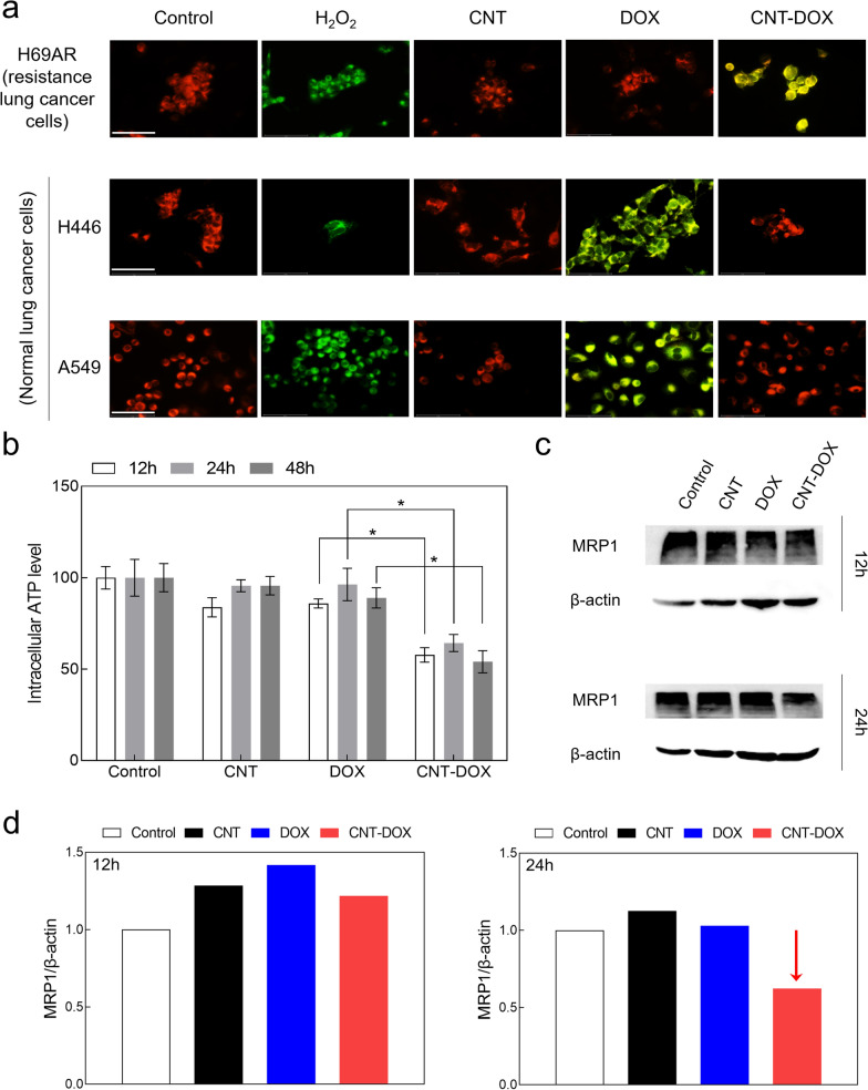
Fig. 7.
Changes in mitochondrial membrane potential (ΔΨm) and reduction of efflux pump. a Confocal images of H69AR showing the JC-1 staining that the depolarized mitochondria (J-monomer, green) were only observed on CNT-DOX treated group and polarized mitochondria (J-aggregate, red) membrane potentials were found on DOX after 24 h. As a positive control, hydrogen peroxide (H2O2) was used. Specifically, CNT-DOX significantly influenced mitochondrial membrane potential in multidrug-resistant lung cancer cells (i.e., H69AR), whereas normal lung cancer cells (i.e., A549 and H446) did not show any notable changes in membrane potential. The scale bar is 75 μm. b Relative intracellular ATP levels in H69AR cells treated with CNT, DOX, and CNT-DOX for the indicated times (12, 24, and 48 h). c Western blot analysis of mrp-1 expression in H69AR cells treated with the nanodrugs (12 and 24 h). d Bar graphs illustrating mrp-1 protein levels quantified by western blot in H69AR cells after drug treatment. Decrease of mrp-1 was only observed on CNT-DOX treated group after 24 h. All protein levels were normalized to the b-actin protein level. All data represent the mean ± SEM (n = 6). *p < 0.05

