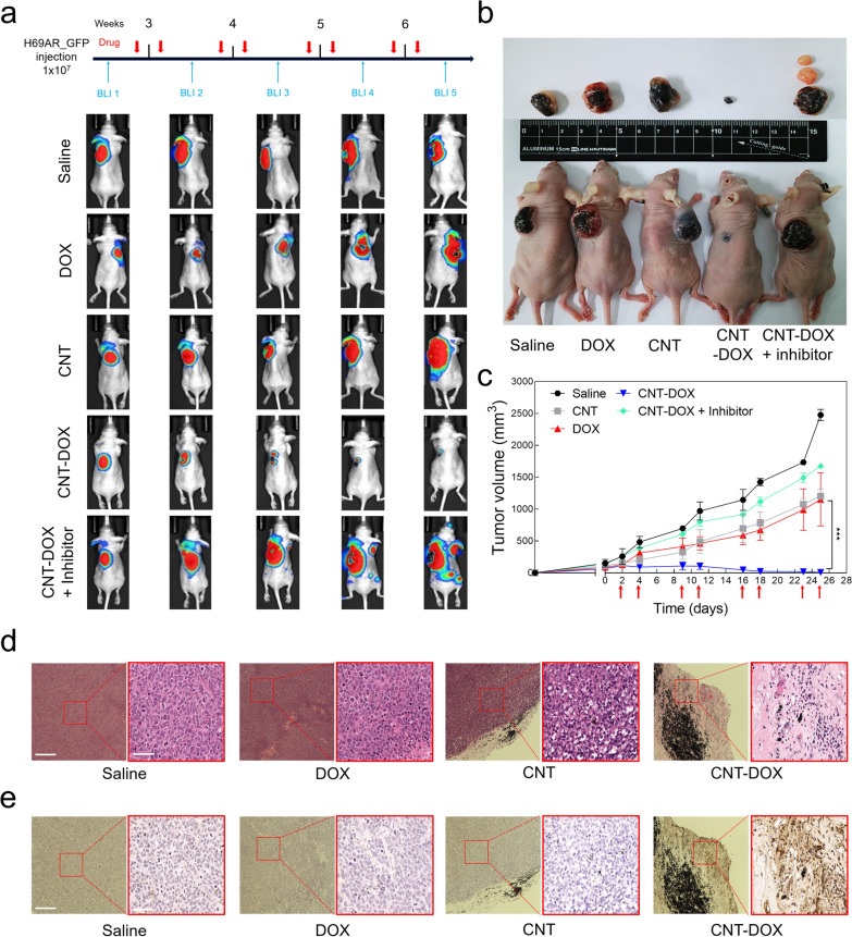
Fig. 8.
Antitumor efficacy of nanodrug on xenograft model mice. a BLI of luciferase expression in H69ARflu-GFP tumor-bearing mice after treatment with PBS, DOX (5 mg/kg), CNT (5 mg/kg), CNT-DOX (5 mg/kg), and CNT-DOX with caveolin inhibitor (5 mg/kg). Red arrow indicates the day of drug injection. b Photographs of H69AR tumor-bearing mice and tumor tissues in week four after drug injection. The smallest tumors with the CNT-DOX treatment are indicated by red arrows. c After the tumor size reached 100 mm3, drugs were injected twice per week for 4 weeks, and tumor size was measured. The therapeutic effect of CNT-DOX on a xenograft mouse model is shown. d Representative images of Hematoxylin and eosin (H&E) staining from H69AR tumor tissues treated with PBS, DOX (5 mg/kg), CNT (5 mg/kg), and CNT-DOX (5 mg/kg). Scale bar of the low- and high-resolution image is 275 μm and 75 μm, respectively. e Terminal deoxynucleotidyl transferase dUTP nick end labeling (TUNEL) staining from H69AR tumor tissues clearly exhibited greater apoptosis on CNT-DOX than on the other groups. Scale bar of the low- and high-resolution image is 275 μm and 75 μm. All data are presented as mean ± SEM (n = 5). ***p < 0.001

