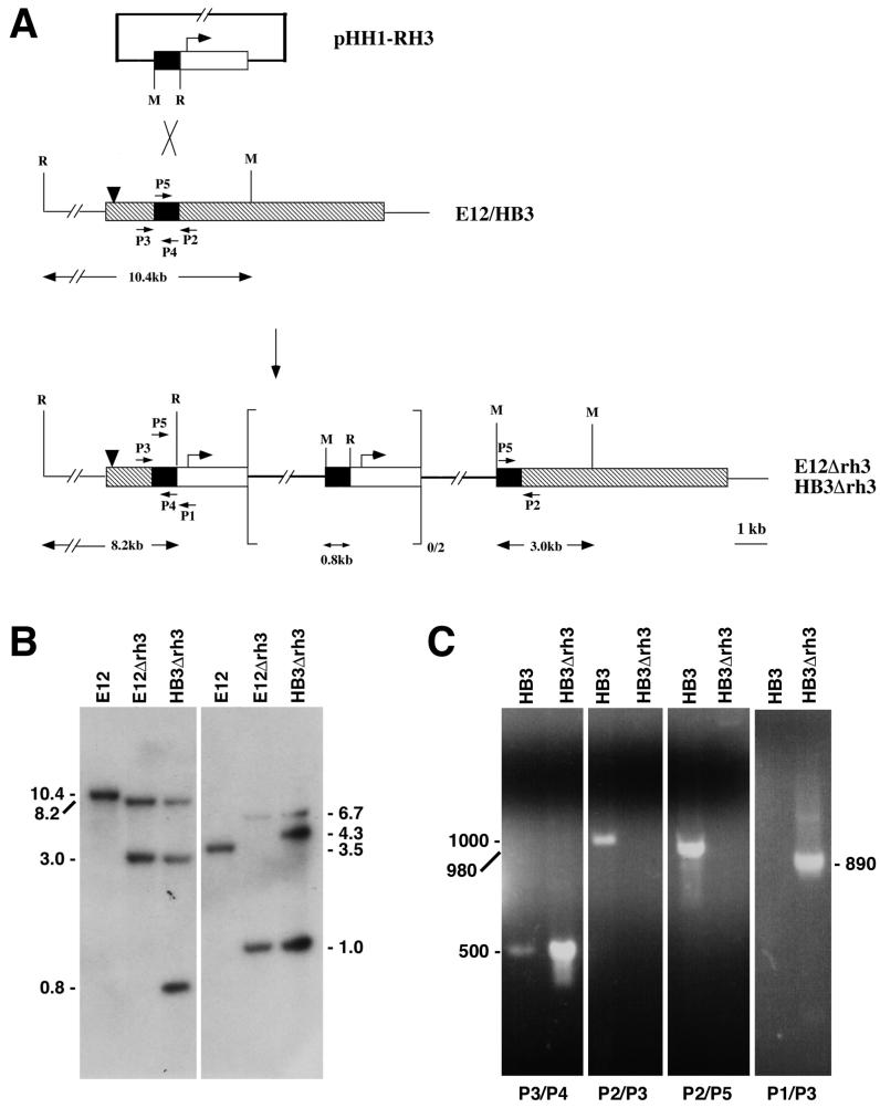
FIG. 4.
PfRH3 is transcribed and can be disrupted in 2 distinct parasite lines. (A) Schematic diagram of the PfRH3 locus in the E12Δrh3 and HB3Δrh3 parasites. The plasmid construct, pHH1-RH3, contains a portion of PfRH3 approximately one-quarter of the way into the gene (solid black shading), which has been cloned in the same direction as the WR99210 selection cassette (no shading). The remainder of PfRH3 in the E12 and HB3 parasites is shown as diagonal hatching. The pGEM vector backbone is shown as the thick black line. The cross indicates the region where homologous integration has occurred. The downward-pointing arrow in the PfRH3 gene represents the single intron. The restriction sites shown are MfeI (M) and EcoRI (R). The cloned parasite line E12Δrh3 has no copies, while the HB3Δrh3 parasite has two copies of the episome integrated at the PfRH3 locus. (B) Southern analysis of untransfected and PfRH3-disrupted parasites. The left panel shows genomic DNAs digested with EcoRI and MfeI, while the right panel shows a BsrGI/XbaI double digest. The Southern blot was probed with a 465-bp PfRH3 fragment within the portion used for the gene disruption. The fragment was amplified using the primers 5′-CTGAAAAGTGTTTTTCGG-3′ and 5′-ATTTATATTATCCATCTTTG-3′. (C) Transcriptional analysis of the PfRH3 locus in HB3Δrh3 parasites. Synchronised late schizont stages from untransfected HB3 and HB3Δrh3 parasites were used for mRNA purification and conversion to cDNA. Primers used to ampify the cDNAs are labeled P1 to P5. Primer pair combinations used for each cDNA are shown below the panels. P1 is a primer within the calmodulin promoter with the sequence 5′-GGTTAACAAAGAAGAAGCTCAGAG-3′. P2 and P3 are from the PfRH3 gene outside the region used for disruption. The P2 sequence is 5′-CTTGATTTAAACTTTTTAGAGGATC-3′, and the P3 sequence is 5′-AAGAATATAACATCTAAAACT-3′. P4 and P5 primers are both within the PfRH3 gene fragment used for disruption. The P4 sequence is 5′-ATTTATATTATCCATCTTTG-3′, and the P5 sequence is 5′-CTGAAAAGTGTTTTTCGG-3′.

