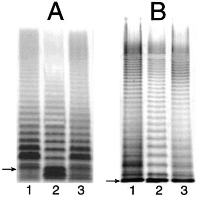
FIG. 2.
Silver-stained SDS-polyacrylamide gels showing the LPS profiles of strains ATCC 14028 (lane 1), CWG304 (lane 2), and CWG304 complemented with the waaP open reading frame from E. coli F470 (lane 3). Samples were run on a 10 to 20% Tricine SDS-polyacrylamide gel (A) and on a standard SDS–12% polyacrylamide gel (B). The gel system in panel A gives better resolution of low-molecular-weight LPS (i.e., LPS lacking O antigen), while the standard gel system shows that the modality of O-antigen expression is unaffected in the mutant strain. The migration of Ra-LPS (lipid A and complete core) in each gel system is indicated by an arrow. Note that Ra-LPS comigrates with LPS molecules with one O-antigen repeat in the gel system in panel B, so the amount of free (uncapped) lipid A-core is misleading. The extent of capping is clear in panel A.

