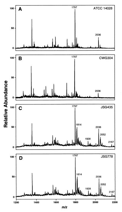
FIG. 4.
Characterization of structural modifications of S. enterica lipid A by negative-ion MALDI-TOF mass spectrometry. All values given are average masses rounded to the nearest whole number for singly charged, deprotonated molecules [M − H]−. (A) ATCC 14028 (parent) lipid A, with the major signal representing the hexa-acylated form (at m/z 1797). The hepta-acylated form containing palmitate (at m/z 2036) is indicated. (B) CWG304 (waaP::aacC1) lipid A, showing ions at m/z 1797 and 2036 as described above. (C) JSG435 (pmrA505) lipid A, showing the hexa- and hepta-acylated forms (as described above), as well as modification by the addition of aminoarabinose (m/z 1928 and 2167, respectively) or by the addition of a hydroxyl group (m/z 1814 and 2052, respectively). (D) JSG778 (pmrA505 waaP::aacC1) lipid A, showing ions at m/z 1797, 1814, 1928, 2036, 2052, and 2167 as described above.

