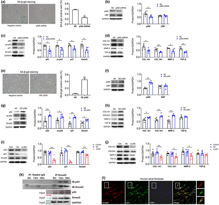
FIGURE 3.

p300‐mediated atrial fibroblast senescence and fibrosis signals through p53/Smad3 pathway in human atrial fibroblasts. (a‐d) Representative SA‐β‐gal staining of senescent human atrial fibroblasts (p11) treated with or without p300shRNA. Scale bars, 100 μm. Representative immunoblots and densitometric analysis of p300/CBP, senescence, and fibrosis‐associated proteins in aged human atrial fibroblasts (P11) treated with p300 shRNA. (e‐h) Representative SA‐β‐gal staining of young cells (p3) treated with or without p300 overexpression. Scale bars, 100 μm. Representative immunoblots and densitometric analysis of p300/CBP, senescence, and fibrosis‐associated proteins in young human atrial fibroblasts (P3) with p300 overexpression. (i and j) Representative immunoblots and densitometric analysis of p53/p21, Smad3, and fibrosis‐associated proteins in aged HAFs (P11) treated with p53 siRNA. *p < 0.05, **p < 0.01; data are mean ± SEM. (k) Co‐immunoprecipitation of p53 with Smad3 in atrial tissue from 5 m, 13 m, and 18 m mice. Immunoblots for p53 (top) and Smad3 (bottom) from samples exposed to protein a/G beads coated with rabbit IgG (negative control) or p53 antibody. (l) Co‐localization of p53 with Smad3 confirmed by confocal microscopy. Human atrial fibroblasts were co‐stained with anti‐p53 antibody, anti‐Smad3, and DAPI. Merged images of p53 (green), Smad3 (red), and DPAI (blue) are shown in the insert. Scale bar represents 100 μm.
