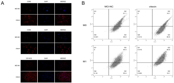
Figure 1. Polarization and identification of M1 macrophages.
(A) Immunofluorescence test determined M1-type macrophages using M1 macrophage surface markers CD80, CD86, and CD16/32. (B) Flow cytometry detection of M0 macrophage polarization results showed that M0 macrophages were polarized into M1 macrophages under the induction of drug stimulation.

