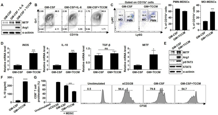
Figure 2.
Induction of BM-MDSC activation and MITF expression by TCCM. BM cells were obtained from the femurs of Balb/c mice, and then lymphocytes were depleted. The cells were cultured in fresh medium with 10 ng/mL GM-CSF in the absence or presence of 10 ng/mL IL-6 or 30% TCCM for 96 hours. (A) MITF and Arg1 in BM-MDSCs were determined by western blot analysis. (B) BM-MDSCs were stained with CD11b and Gr1 antibodies and analyzed by flow cytometry. (C) Cells were stained with CD11b, Ly6C, and Ly6G antibodies and analyzed by flow cytometry. (D) The mRNA expression of iNOS, IL-10, TGF-β, and MITF in BM-MDSCs was measured by real-time PCR. (E) MITF, pSTAT3, and Arg1 were evaluated by western blot analysis. (F) BM cells were treated with TCCM for 96 hours and then cultured in serum-free medium. After 48 hours, the supernatant was collected and used to detect IL-10 by ELISA. (G) Splenic CD3+ T cells were labeled with 2.5 µM CFSE. CFSE-labeled T cells were stimulated with 3 µg/mL plate-bound anti-CD3 mAb and 1 µg/mL soluble anti-CD28 mAb for 2 hours. BM-MDSCs were cocultured with CFSE-labeled CD3+ T cells for 72 hours, and then CD8+ T-cell proliferation was measured by flow cytometry at an MDSC:T-cell ratio of 1:2. All experiments were independently repeated at least three times. *P<0.05, ***P<0.001, ****P<0.0001. Arg1, arginase 1; BM, bone marrow; GM, granulocyte–macrophage; GM-CSF, granulocyte–macrophage colony-stimulating factor; IL, interleukin; iNOS, inducible nitric oxide synthase; MDSC, myeloid-derived suppressor cell; MITF, microphthalmia-associated transcription factor; MO, monocytic; PMN, polymorphonuclear; TCCM, tumor cell-conditioned medium; TGF-β, transforming growth factor beta.

