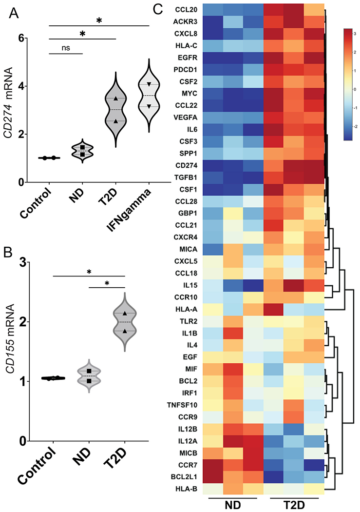
Fig. 3.

T2D plasma exosomes upregulate genes that encode immune checkpoint ligands in prostate cancer cells. Plasma exosomes from T2D subjects (▴) upregulated CD274 (A) and CD155 (B) mRNA expression in DU145 cells after 2 days, compared to ND control (∎). Treatment with IFN-γ (5 ng/mL) was the positive control for CD274 induction (▾), as we previously published [53]. Treatment with growth media exosomes was the negative control (●). Data in A and B were obtained from two biological replicates of ND and T2D, and each biological replicate was conducted in three technical replicates. Data were analyzed by two-way ANOVA with statistical significance presented as: *, P μ 0.05; ns, not significant. (C) Hierarchical clustering of genes involved in inflammation and cancer immune crosstalk, analyzed by commercial PCR array. DU145 cells were treated with plasma exosomes from three T2D subjects or three ND subjects. Equal numbers of exosomes (109) from each sample were used. The heatmap of the PCR array result was calculated by hierarchical clustering. Scale bar (right) shows fold change, with red color to identify upregulation and blue color to identify downregulation. (ND, non-diabetic; T2D, Type 2 diabetic; Control, media-only exosomes; IFNgamma, interferon-γ). (For interpretation of the references to color in this figure legend, the reader is referred to the Web version of this article.)
