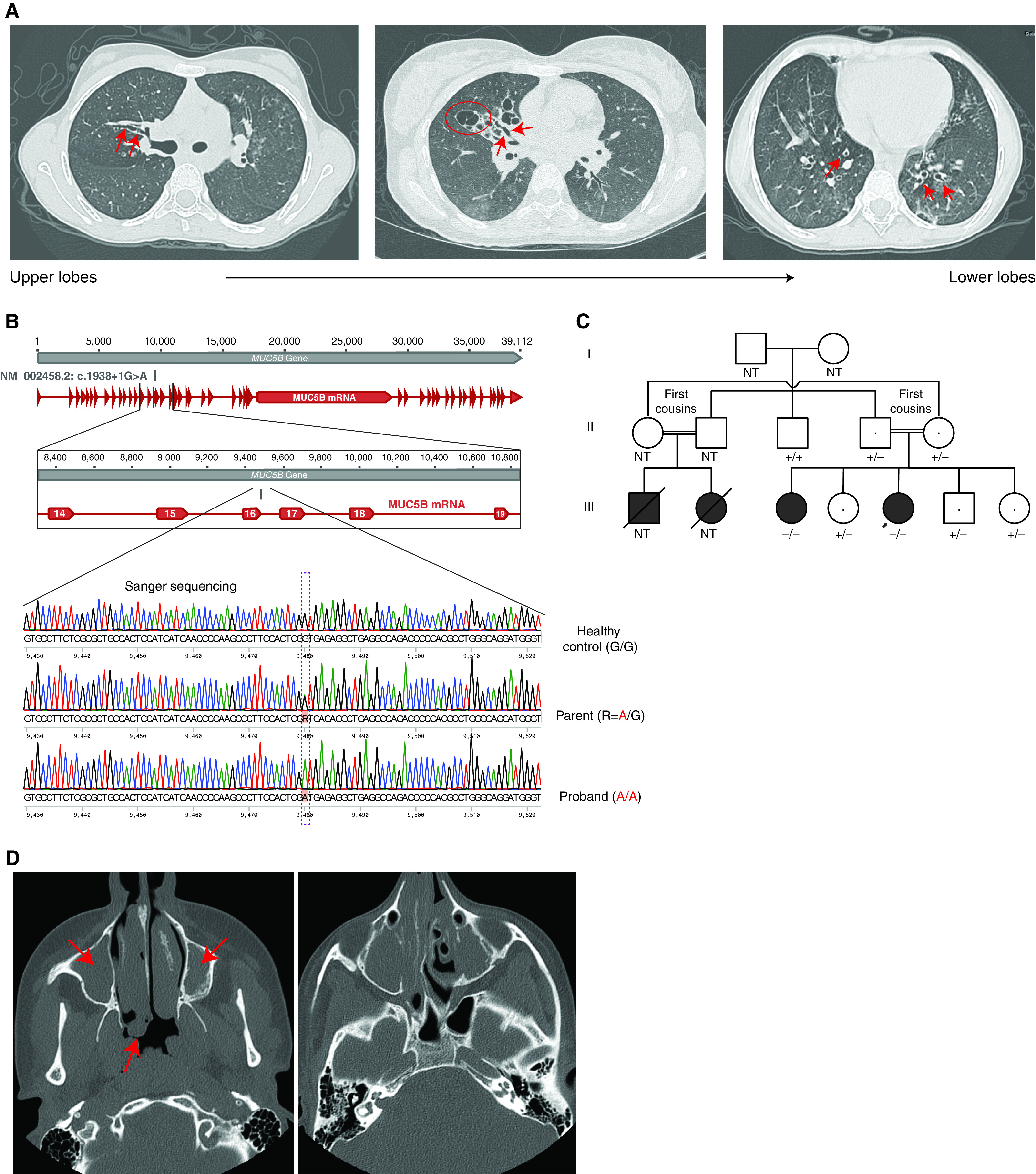
Figure 1.

Phenotype and genotype information for the study participants. (A) High-resolution computed tomography (CT) images from the proband at age 10 years showing cylindrical (red arrows) and associated saccular bronchiectasis (e.g., red circle), most severe in the right upper and middle lobes. (B) Sanger sequencing chromatograms for an individual homozygous for the MUC5B reference allele (healthy control subject), an individual heterozygous for the MUC5B alternate allele (parent of proband), and an individual homozygous for the MUC5B alternate allele (proband). (C) Pedigree for the index family. −/− = homozygous for the alternate allele; +/− = heterozygous for the alternate allele; +/+ = homozygous for the reference allele. The arrow indicates the proband. (D) Volumetric axial sinus CT image from the proband’s affected sister at age 15 years showing extensive bilateral nasal polyposis (red arrows). NT = not tested.
