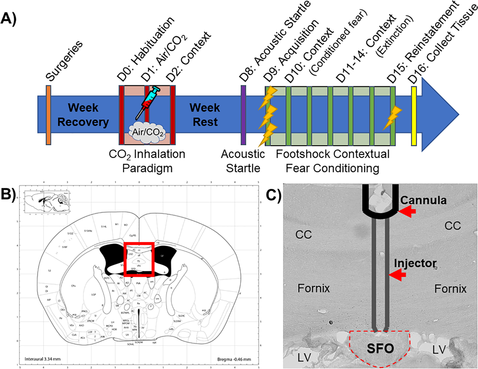
Fig. 1.

A) Schematic overview of the experimental timeline. Mice received cannulations targeting the subfornical organ (SFO) and were allowed to recover for 1 week before undergoing the CO2 inhalation paradigm (see text for details). Mice received a single IL-1RA or vehicle infusion into the SFO 30 min prior to either CO2 or air inhalation then were re-exposed to inhalation context the next day. Mice were left undisturbed for a week, after which they were exposed to the acoustic startle test followed by a contextual fear conditioning paradigm the next day (see text for details). Tissue was collected the day after all behavioral testing was completed. B) Image from brain atlas (Paxinos and Watson) showing location of the SFO (bregma. AP −0.48 mm, DV 2.4 mm, ML 0 mm). C) A representative image showing a SFO “hit” with injector tracts into the dorsal SFO. D = Day; CC = corpus callosum; LV = lateral ventricle.
