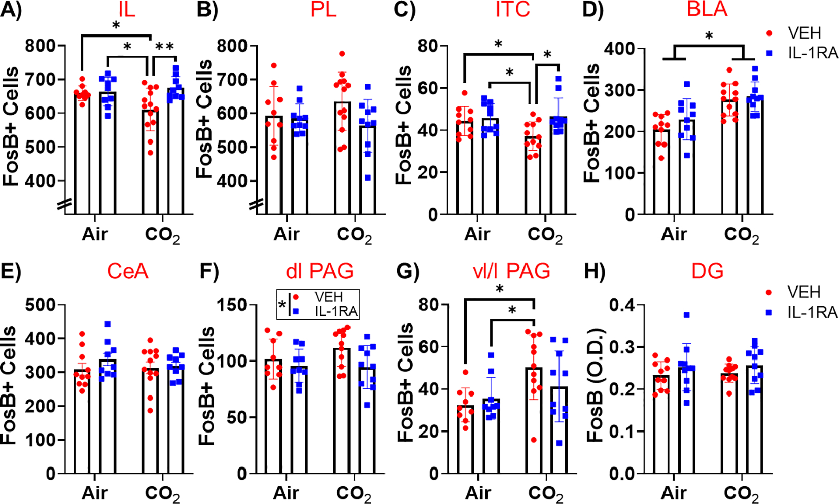
Fig. 4.

Post behavior regional ΔFosB + cell counts in CO2– or air- exposed mice treated with vehicle or IL-1RA. (A) Within infralimbic cortex (IL), IL-1RA restored the CO2 attenuation of FosB + cell counts. (B) Neither CO2 or IL-1RA affected FosB + cell counts in the prelimbic cortex (PL). (C) Within the intercalated cells of the amygdala (ITC), IL-1RA restored the CO2 attenuation of FosB + cell counts. (D) CO2 inhalation increased basolateral amygdala (BLA) FosB + cell counts, but there was no effect of IL-1RA. (E) Neither CO2 or IL-1RA affected FosB + cell counts in the central amygdala (CeA). (F) IL-1RA reduced FosB + cell counts in the dorsolateral periaqueductal grey (dl PAG). (G) CO2 inhalation increased FosB + cell counts in the ventrolateral/lateral PAG (vl/l PAG) and CO2/VEH mice showed increased FosB + cell counts compared to Air/VEH and Air/IL-1RA mice. (H) Neither CO2 or IL-1RA affected FosB + cell counts in the dentate gyrus of the hippocampus (DG). Data are mean ± SEM. *p < 0.05 **p < 0.01 (n = 8–13).
