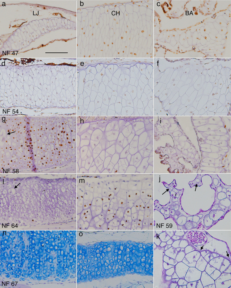
Fig 4. Representative sections of lower jaw (LJ), ceratohyal (CH) and branchial arch (BA) cartilages showing chondrocyte size, shape, arrangement, and matrix at different stages.
(a-i, l-o) frontal, BrdU-labeled, hematoxylin or alcian blue-counterstained sections through middle portions of left LJ, CH and second ceratobranchial cartilages at NF 47, 54, 58, 64, and 67 (right is to the midline, up is anterior or lateral); arrows in g and l indicate large chondrocytes in the outer (more lateral) half of the cartilage. (j and k) transverse, resin-embedded, H&E-stained sections of branchial arch cartilages at NF 59. J shows an ornate process containing round chondrocytes of different sizes (arrows point to the smallest), capillaries, interstitial space, epithelia, and no perichondrium. K shows the transition from ceratobranchial base (lower right) to vertical rod (upper left), which is marked by a decrease in cell size, change from polygonal to more rounded cell outlines, and absence of perichondral cells (arrows) in the vertical rod. Scale bar is 0.1 mm.

