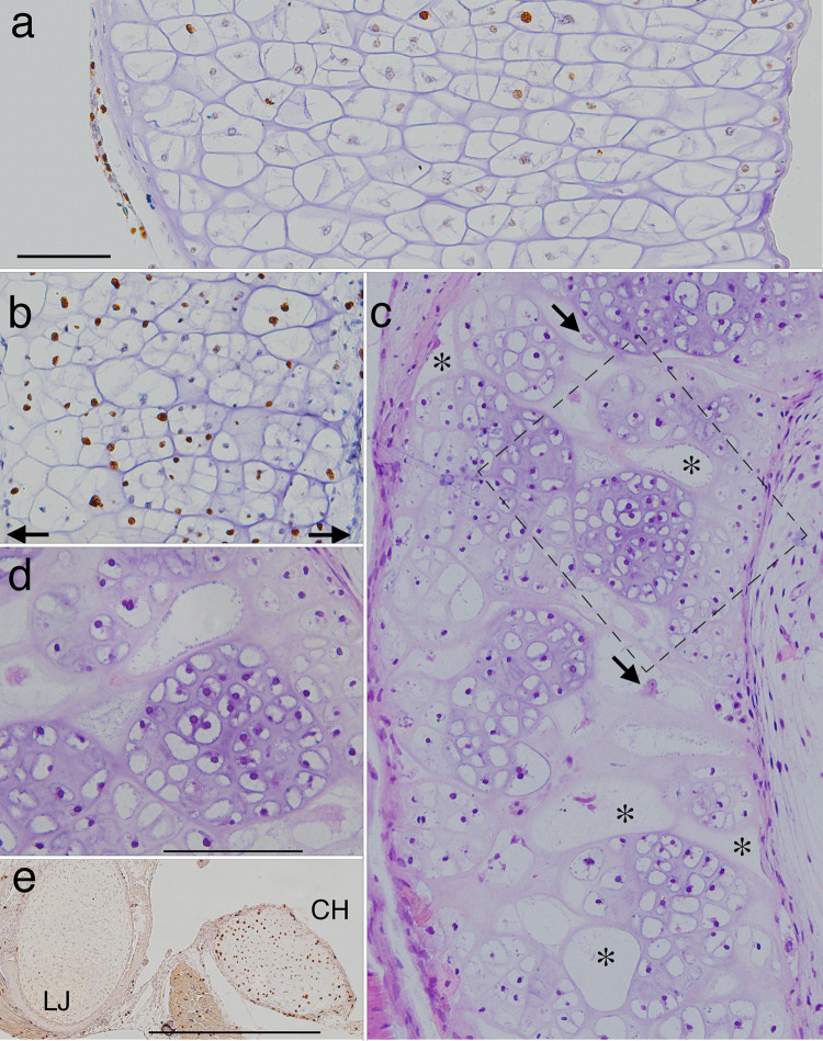
Fig 11. Sections through the proximal half of left ceratohyals showing metamorphic changes in cartilage histology, shape and cell features.
Left is to the outside of the head and up is anterior in a-c and dorsal in e. (a and b) frontal, BrdU-labeled, hematoxylin-counterstained sections at late larval (NF 58) and late metamorphic (NF 64) stages respectively (arrows in b indicate the edges of the cartilage). (c) a frontal, resin-embedded, H&E stained section through the same region as a and b soon after metamorphosis (NF 66+). (d) a close up of the rectangular region outlined in c. (e) a transverse, BrdU-labeled, hematoxylin-counterstained section through the left lower jaw (LJ) and ceratohyal (CH) at NF 66. The transition from NF 58 to 64 (a to b) is marked by narrowing of the cartilage and most large chondrocytes acquiring more daughter nuclei and cells within their original perimeters. The transition from NF 64 to 66+ (b to c) is marked by the emergence of large, largely empty cell lacunae (*) that are interspersed across the width of the cartilage with discrete, equantly shaped cell clusters, and smaller lacunae that appear to contain cellular debris (arrows). While some lacunae resemble cluster outlines in b, others are transversely elongated with oval or more irregular shapes that generally conform to the curvature of adjacent cell clusters. The cell clusters contain many, small chondrocytes that have spherical nuclei and are separated by more matrix than the chondrocytes at NF 58. E shows high BrdU labeling in the ceratohyal, but not the lower jaw, at NF 66. Scale bars for a-d and e are 0.1 and 0.5 mm respectively.

