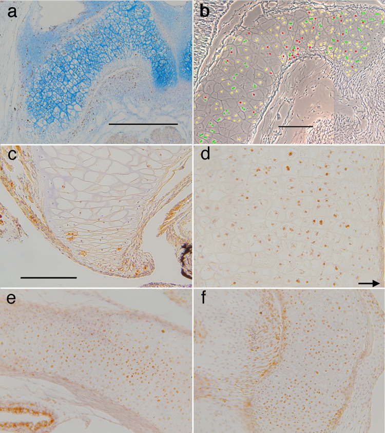
Fig 12. Frontal sections through left ceratohyals showing distributions of cell division and death labels.
Anterior is up for a-d, and to the right for e-f. (a) BrdU labeled chondrocytes and alcian blue staining of matrix persist across the width of the cartilage to late metamorphosis (NF 65). (b) a phase micrograph at NF 63 showing the locations of DAPI-stained chondrocyte nuclei that appear to have recently divided (green), have undergone nuclear fragmentation (red), and be in interphase (yellow); see Fig 2B for scoring criteria. Recently divided nuclei are interspersed with ones exhibiting nuclear fragmentation. (c) capase labeling of a few peripheral chondrocytes in the distal tip at NF 63. (d-f) PCNA labeling of chondrocytes is strongest in the cartilage center at NF 62/3 (d) and 65 (e-f); arrow in d indicates the medial edge of the cartilage; scale bars for a, b and c-f are all 0.2 mm.

