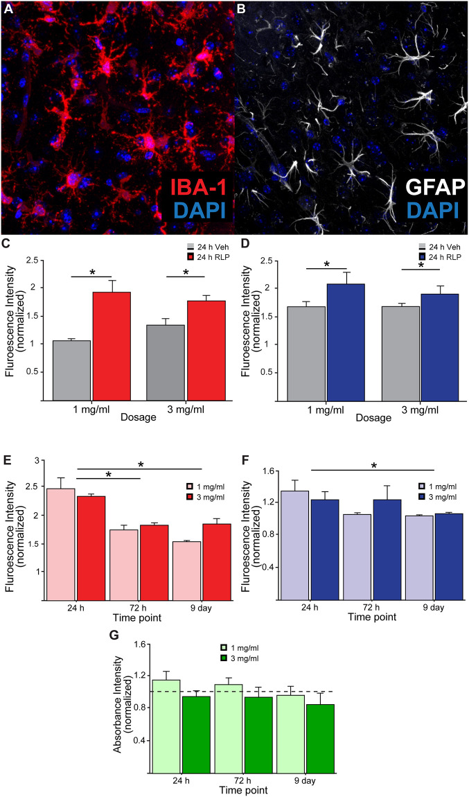
Fig 5. Quantifying immune cell presence adjacent to radioluminescent particle injections.
A) Representative image of IBA-1+ microglia and B) GFAP+ astrocytes near RLP injection site. C) Fluorescence intensity of IBA-1 and D) GFAP staining in vehicle (saline) versus RLP-injected M2 following injections of 1mg/ml and 3mg/ml in mice sacrificed 24 hours after injection. E) Total fluorescence intensity of IBA-1 and F) GFAP in RLP-injected M2 (normalized to vehicle-treated contralateral hemisphere) for animals receiving 1 and 3 mg/ml concentrations of RLPs 24 hours, 72 hours, and 9 days post-injection. (G) In vitro Alamar blue assay revealed no significant changes in cell viability of cultured neurons following RLP exposures at multiple timepoints compared to untreated controls (dashed line at “1”).

