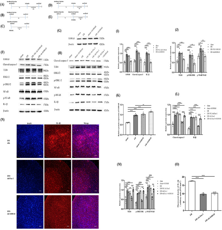
FIGURE 6.

Fra‐1/S100A8‐mediated TLR4 signaling promotes ERK1/2 and NF‐κB pathways activation and IL‐1β production after spinal I/R. (A) Schematic of the experimental design shows the timeline for grouping rats. The rats were intrathecally injected sh‐S100A8 7 days before I/R induction. After 48 h of I/R, tissue samples were collected. (B) The rats were intrathecally injected rS100A8, and tissue samples were collected 48 h later. (C) The rats were intrathecally injected rS100A8 before I/R induction. After 48 h of I/R, tissue samples were collected. (D) The rats were intrathecally injected sh‐Fra‐1 7 days before I/R induction. After 48 h of I/R, tissue samples were collected. (E) The rats were intrathecally injected sh‐Fra‐1 7 days before administration of rS100A8 and intrathecally injected rS100A8 before I/R induction. After 48 h of I/R, tissue samples were collected. (F‐M) Western blot‐assisted analysis and quantification of the protein levels of S100A8, cleaved caspase‐3, TLR4, p‐ERK/ERK, p‐NF‐κB/NF‐κB and IL‐1β (n = 5 rats/group). (N) Immunofluorescence (IF) assays of IL‐1β expression in different groups were performed (n = 3 rats/group). Scale bar =100 μm. (O) Quantification of the IL‐1β fluorescence intensity (INT/mm2). All data represent the mean ± SEM. *p < 0.05, **p < 0.01, ***p < 0.001, and ****p < 0.0001.
