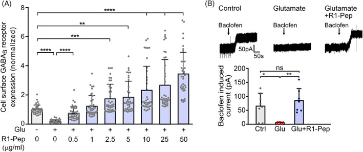
FIGURE 2.

R1‐Pep dose‐dependently increased cell surface GABAB receptors after glutamate treatment and restored normal GABAB receptor function. Cultures were stressed for 1 h with 50 μM glutamate and thereafter treated with R1‐Pep. The neurons were then incubated for 16 h with the peptide and analyzed. (A) Neurons were incubated overnight with increasing concentrations of R1‐Pep and stained for surface expression of GABAB receptors using GABAB2 antibodies. Signals were normalized to no Pep/no Glu control. N = 38 neurons per condition from three independent experiments. One‐way ANOVA, Dunnett's T3 multiple comparison test (**, p < 0.01; ***, p < 0.001; ****, p < 0.0001). (B) R1‐Pep (10 μg/ml) restored normal GABAB receptor function. Baclofen‐induced currents were measured in glutamate‐stressed (Glu) and unstressed (Ctrl) neurons using whole‐cell patch‐clamp recordings. N = 8 (Ctrl), 6 (Glu), and 7 (Glu + R1‐Pep) neurons. One‐way ANOVA, Tukey's multiple comparison test (ns, p > 0.05; *, p < 0.05; **, p < 0.01).
