
FIGURE 7.
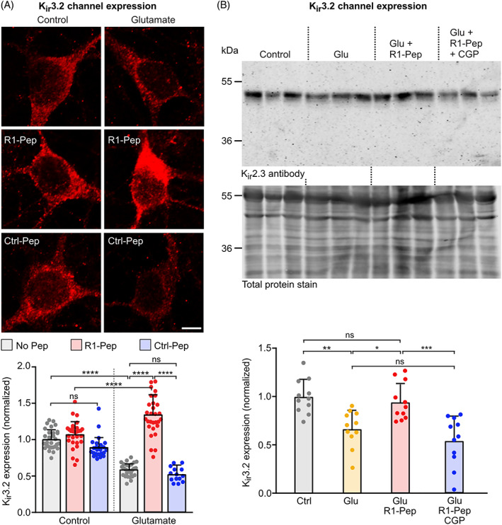
R1‐Pep treatment reduced aberrant expression of Kir3.2 channels after glutamate stress. Glutamate‐stressed neurons were treated for 16 h with R1‐Pep (10 μg/ml) or Ctrl‐Pep (10 μg/ml) and then stained with Kir3.2 antibodies. (A) Immunofluorescence staining: top, representative images (scale bar: 5 μm); bottom, quantification of fluorescence intensities. N = 30 neurons per condition from two independent experiments. Signals were normalized to no Pep control. Two‐way ANOVA with Tukey's multiple comparison test (ns, p > 0.05; ****, p < 0.0001). (B) Western blotting: top panel, staining for Kir3.2 channels; bottom panel, staining for total protein used for normalization. Control, untreated cultures; Glu, glutamate‐stressed cultures; Glu + R1‐Pep, glutamate‐stressed cultures treated with R1‐Pep; Glu + R1‐Pep + CGP, glutamate‐stressed cultures treated with R1‐Pep and the GABAB receptor antagonist CGP 56999. Signals were normalized to control. N = 11 cultures per condition from four independent neuron preparations. One‐way ANOVA with Tukey's multiple comparison test (ns, p > 0.05; *, p < 0.05; **, p < 0.01 ***, p < 0.0005).