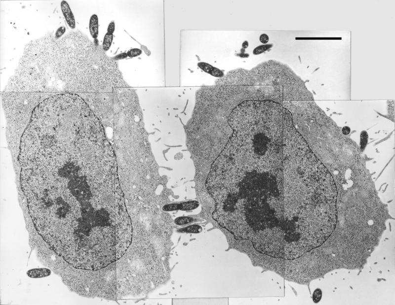
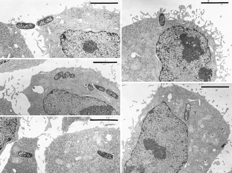
FIG. 2.
Typical electron microscopic images of Pla+ and Pla− Y. pestis interactions with HeLa cells at the time of harvest in a 60-min invasion assay. Prior to addition of the glutaraldehyde prefixative, the cultures for electron microscopy were handled exactly as in our optimized invasion assay. In parallel, replicate cultures were assayed for invasion by the GM protection assay. For the experiment depicted, the Pla+ Y. pestis showed 33% invasion, whereas the Pla− strain showed 3.2% invasion. (Left panel) Pla−. Five adjacent frames are combined into one montage. (Right panel) Pla+. Five nonadjacent frames are presented. Bars, 5 μm.


