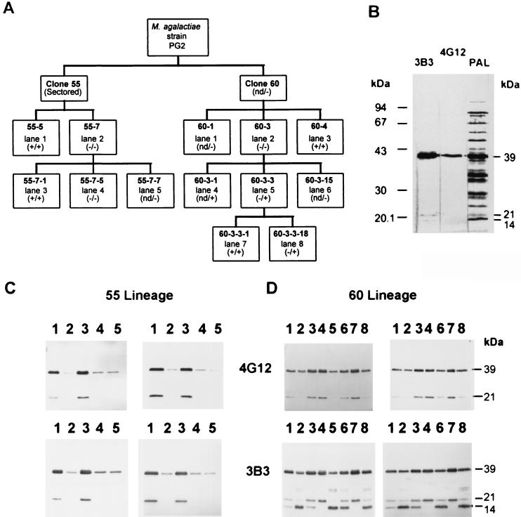
FIG. 1.
(A) Outline showing clones of the 55 (from clone 55) and 60 (from clone 60) lineages, derived from M. agalactiae type strain PG2 based on colony immunostaining positive (+) or negative (−) with MAb 3B3. These clones were also immunostained at the colony level with MAb 4G12 except for those marked “nd” (not determined). (−/+) indicates 4G12-negative and 3B3-positive colonies; (−/−) indicates 4G12- and 3B3-negative colonies. (B) Western blot analysis of Triton X-114-phase material from the parental M. agalactiae type strain PG2 using 3B3, 4G12, or polyclonal anti-M. agalactiae sheep serum (PAL) as indicated above each lane. (C and D) Western blot analysis of colony clones from the 55 (C) and 60 (D) lineages. Whole-cell lysates (left panels) and cell material partitioned into the Triton X-114 detergent phase (right panels) were immunostained with 4G12 (top panels) and 3B3 (bottom panels). Lanes are labeled according to the lane number shown in panel A for the corresponding lineage.

