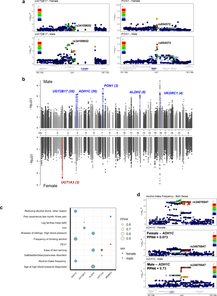
Fig. 2. Sex differences in the genetic regulation of gene expression in human liver in part mediate human complex traits.
a Example of sex-differentiated/specific cis-DMET eQTLs in both sexes for UGT2B17 (left panels) and PON1 (right panels) in liver. b Manhattan plot of sex-stratified cis-eQTLs in DMET regions in human liver. Sex-differentiated/specific cis-eQTLs and their corresponding genes are labeled (blue: Male, red: Female) and parentheses indicates the number of cis-eQTLs. Highlighted genes are statistically significant after multiple testing correction (FDR < 0.1). c Colocalization of GWAS traits with sex-differentiated/specific cis-eQTLs for DMET genes. PPH4 values are represented by the size of circles. Only colocalizations with PPH4 > 0.5 in one sex are shown (females: red; males: blue). d LocusZoom plots for alcohol intake frequency at the ADH1C locus. The top panels illustrate the results from the sex-combined GWAS, while female-only and male-only cis-eQTLs are shown in the middle and the bottom panels. rs34076947 is a male-specific ADH1C cis-eQTL in liver. In (a) and (d), linkage disequilibrium between loci is quantified by the squared Pearson coefficient of correlation (r2). In (a, b, d), association testing was performed using a linear regression model by FastQTL.

