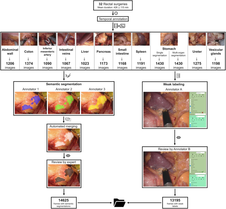
Fig. 1.
Overview of the data acquisition and validation process. Based on temporal annotations of 32 rectal resections, three independent annotators semantically segmented every single image with regard to the pixel-wise location of the respective organ. These segmentations were merged and individual segmentations were reviewed alongside the merged segmentation by a physician with considerable experience in minimally-invasive surgery, resulting in the final pixel-wise segmentation (left panel). Moreover, every single image was classified with regard to the visibility of all individual anatomical structures of interest by one annotator and independently reviewed (right panel).

