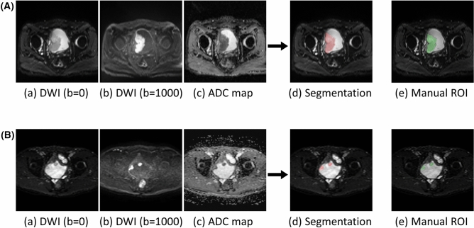
Figure 4.
Two representative cases of automatic segmentation of bladder cancer in the test dataset (case 1 (A); case 2 (B)). (a) Diffusion weighted image with b = 0 s/mm2, (b) diffusion weighted image with b = 1000 s/mm2, (c) apparent diffusion coefficient map, (d) results of automatic segmentation overlayed on diffusion weighted image with b = 0 s/mm2, (e) manual region of interest for the reference standard. The large tumor was almost perfectly segmented in case 1 (Dice similarity coefficient = 0.95), and the two distant tumors were well segmented in case 2 (Dice similarity coefficient = 0.82).

