. 2023 Jan 15;24(1):32–49. doi: 10.1631/jzus.B2200269

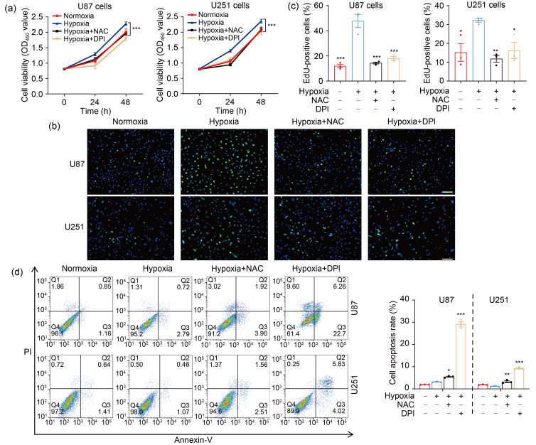
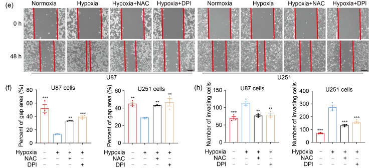
Fig. 2. ROS mediate hypoxia-promoted GBM progression. (a) CCK-8 assay was applied to examine the cell viability at 24 and 48 h. (b, c) EdU analysis was conducted to assess the cell proliferation ability at 48 h (scale bar=50 μm). (d) Flow cytometry was performed to detect the effect of ROS on cell apoptosis at 48 h. (e, f) The cell migration ability was measured by wound healing assay at 48 h (scale bar=200 μm). (g, h) The cell invasion ability was determined by transwell matrigel assay at 48 h (scale bar=100 μm). For all functional trials, the normoxia and hypoxia groups were used as negative and positive control, respectively, and the inhibitor groups were experimental groups. For experimental groups, cells were pretreated using NAC or DPI for 4 h before hypoxic exposure for the indicated time periods. Data are expressed as mean±SEM (n=3). * P<0.05, ** P<0.01, *** P <0.001, vs. hypoxia group. ROS: reactive oxygen species; GBM: glioblastoma; CCK-8: cell counting kit-8; EdU: 5-ethynyl-2'-deoxyuridine; NAC: N-acetyl-L-cysteine; DPI: diphenyleneiodonium chloride; SEM: standard error of the mean; OD450: optical density value at 450 nm; PI: propidium iodide.

graphic file with name JZhejiangUnivSciB-24-1-32-g002a.jpg

graphic file with name JZhejiangUnivSciB-24-1-32-g002b.jpg

graphic file with name JZhejiangUnivSciB-24-1-32-g002c.jpg