. 2023 Jan 15;24(1):32–49. doi: 10.1631/jzus.B2200269

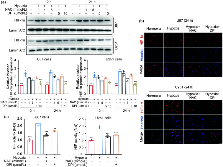
Fig. 3. Hypoxia-induced ROS drive HIF-1α activation in GBM cells. (a) The nuclear expression of HIF-1α protein was detected and quantified by western blotting after cells were incubated in the presence or absence of NAC (1 or 5 mmol/L) and DPI (5 or 10 μmol/L) for 4 h followed by hypoxia for 12 and 24 h. (b) The localization of HIF-1α was assessed by immunostaining in U87 and U251 cells under hypoxia 24 h after NAC (5 mmol/L) and DPI (10 μmol/L) treatment for 4 h (scale bar=25 μm). (c) Luciferase assay was performed to analyze the effect of ROS on HIF-1α activity. Cells that had been transfected using HIF-1α-luciferase plasmid for 20 h were treated with 5 mmol/L NAC or 10 μmol/L DPI for 4 h and then incubated under hypoxic conditions for 24 h. The normoxia group was used as negative control. Data are expressed as mean±SEM (n=3). * P<0.05, ** P<0.01, *** P<0.001, vs. hypoxia group. ROS: reactive oxygen species; HIF-1α: hypoxia-inducible factor-1α; GBM: glioblastoma; NAC: N-acetyl-L-cysteine; DPI: diphenyleneiodonium chloride; SEM: standard error of the mean.

Fig. 3