. 2023 Jan 15;24(1):32–49. doi: 10.1631/jzus.B2200269

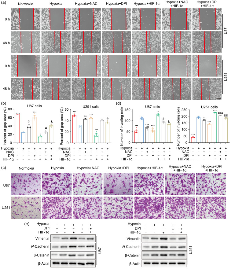
Fig. 4. ROS potentiate cell migration and invasion though HIF-1α signaling pathway in hypoxic GBM cells. (a, b) Scratch wound assay was used to test the effect of HIF-1α on cell migration under hypoxia for 48 h (scale bar=200 μm). (c, d) Matrigel invasion assay was performed to examine the effect of HIF-1α on cell invasion under hypoxia for 48 h (scale bar=100 μm). (e) The expression of EMT-related proteins under hypoxia for 24 h was detected by western blotting. For all experiments, cells were transfected with or without HIF-1α expression plasmid, and then incubated in the presence or absence of NAC or DPI for 4 h before hypoxia treatment for 24 or 48 h. The normoxia group was used as negative control. Data are expressed as mean±SEM (n=3). * P<0.05, ** P<0.01, *** P<0.001, vs. hypoxia group; # P<0.05, ### P<0.001, vs. hypoxia+NAC group; & P<0.05, && P<0.01, vs. hypoxia+DPI group. ROS: reactive oxygen species; HIF-1α: hypoxia-inducible factor-1α; GBM: glioblastoma; EMT: epithelial-mesenchymal transition; NAC: N-acetyl-L-cysteine; DPI: diphenyleneiodonium chloride; SEM: standard error of the mean.

Fig. 4