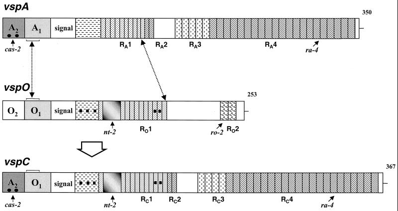
FIG. 7.
Schematic representation of the generation of the vspC gene by an intrachromosomal recombination between vspA and vspO. Structures of vspA, vspO, and vspC are presented schematically by aligned rectangles. Each vsp gene is flanked 5′ by a highly conserved noncoding region composed of two cassettes, labeled 1 and 2 with the letter of the corresponding gene. A block labeled signal represents a highly homologous sequence encoding a lipoprotein signal peptide. In-frame reiterated coding sequences extending from the N terminus to the C terminus of the Vsp proteins and encoding periodic amino acid sequences are shown by differently hatched blocks. Distinctive repetitive domains within each Vsp are labeled with R and the letter of the corresponding vsp gene. Repetitive units present in more than one Vsp molecule are similarly hatched. Numbers on the right indicate the length of each Vsp polypeptide chain. Small black squares denote distinctive nucleotide changes which exist in vspO and vspC but not in vspA, while solid dots represent a distinctive signature present in vspA and vspC but not in vspO. A shaded block represents a 66-bp sequence present in vspO and vspC but absent in vspA. Small labeled arrows mark the locations of the cas-2, ro-2, nt-2, and ra-4 oligonucleotides used as probes in Southern blot experiments. The position of a 34-bp sequence, within cassette 1 of the vspA, vspO, and vspC genes, which served as the putative 5′-end site for the recombination event is marked by a broken arrow and brackets. The 3′ end of the recombination event, which is located within the repetitive domains RA1 and RO1 of the vspA and the vspO genes, is marked by a broken line.

