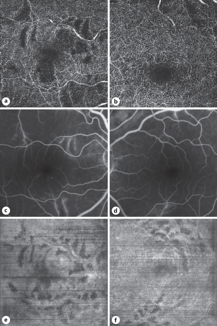
Fig. 2.
En face OCT reconstruction, OCT angiography, and FA. a, b OCT angiography at the level of the deep capillary plexus. The areas devoid of capillaries coincide with the areas of thinning in a and b above, indicating the conjunction of DCP ischemia with the late PAMM-like lesions. c, d Enlarged images of the FA, to facilitate the comparison with the en face reconstructed images and OCT angiography. e, f En face OCT reconstruction at the INL-OPL level with concurrent shadow removal revealing dark areas of INL thinning consistent with late PAMM lesions. FA, fluorescein angiography; OPL, outer plexiform layer.

