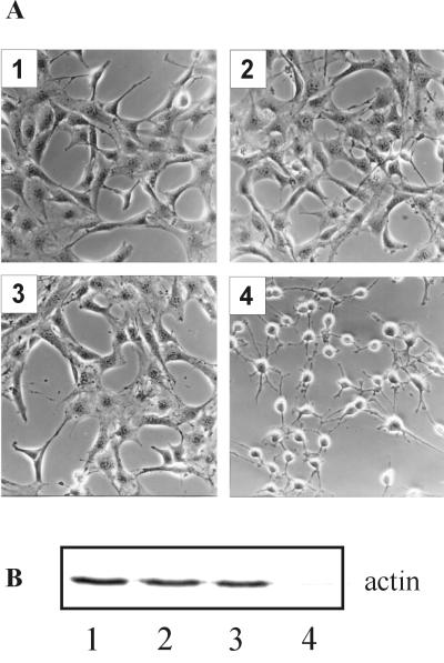
FIG. 2.
Cytotoxic effect of recombinant C2 toxin on NIH 3T3 cells. NIH 3T3 cells were incubated at 37°C in HBSS with C2 toxin components. Three hours after toxin addition cells were photographed (A), and cell lysates were analyzed by subsequent in vitro ADP-ribosylation of actin (B). Cell lysates were incubated with C2I and [32P]NAD. Actin that had not been modified by the toxin pretreatment was [32P]ADP-ribosylated in the in vitro assay and detected by SDS-PAGE and phosphorimaging. (A) Image 1, control cells; image 2, cells treated with 200 ng of C2II per ml plus 100 ng of C2I per ml; image 3, cells treated with 200 ng of C2IIa per ml; image 4, cells treated with 200 ng of C2IIa per ml plus 100 ng of C2I per ml.

