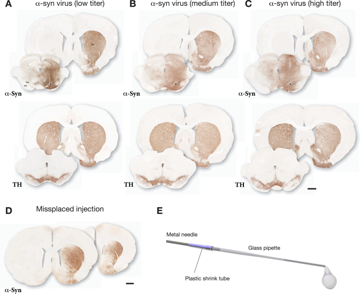
Fig. 1.
Example of the outcome of a pre-test made in order to select a suitable dose for the AAV-α-syn vector. The vector was injected at 3 different doses as a single 3μL deposit in the SN and the brains were processed for immunohistochemistry three weeks later. The extent and distribution of vector-derived α-syn was determined in sections through striatum and SN using antibodies against TH (Sheep ab1542) and human α-syn (Rabbit ab5038 from Millipore). The selected low titer dose illustrated in A showed good spread of AAV-derived human α-syn, covering the medio-lateral extent of the SN, and α-Syn+terminals (expressing α-syn as a result of anterograde transport) was seen to cover the entire medio-lateral extent of the caudate-putamen. This wide-spread distribution of α-syn-expressing terminals indicates that the vast majority of nigral DA neurons (as well as some VTA neurons) had been transduced by the vector. TH immunostaining showed no clearly detectable TH+cell loss, or loss of TH+innervation in the striatum. The medium titer dose illustrated in B (5-fold higher than in A) showed some spread of α-syn into the contralateral side and reduced TH staining in both striatum and SN. At the highest dose illustrated in C (16.5-fold higher than in A) the vector-derived α-syn extended well beyond the ventral midbrain/SN area and spread also across the midline into the contralateral non-injected side, and there was a clear loss of TH+neurons in the SN and TH+innervation in the striatum. For this vector batch the selected dose was 3.6 × 10⁹ gc in 3μL (titer determined by PCR using a probe recognizing the ITR). D shows the uneven expression of α-syn in striatum resulting from a minor displacement of the injection, 0.7 mm lateral to the one shown in A–C. E shows the glass capillary, attached to the 22-gauge metal cannula of a Hamilton syringe, used for the injections. Asterisks mark the injection sites. Scale bars in A–D: 1 mm.

