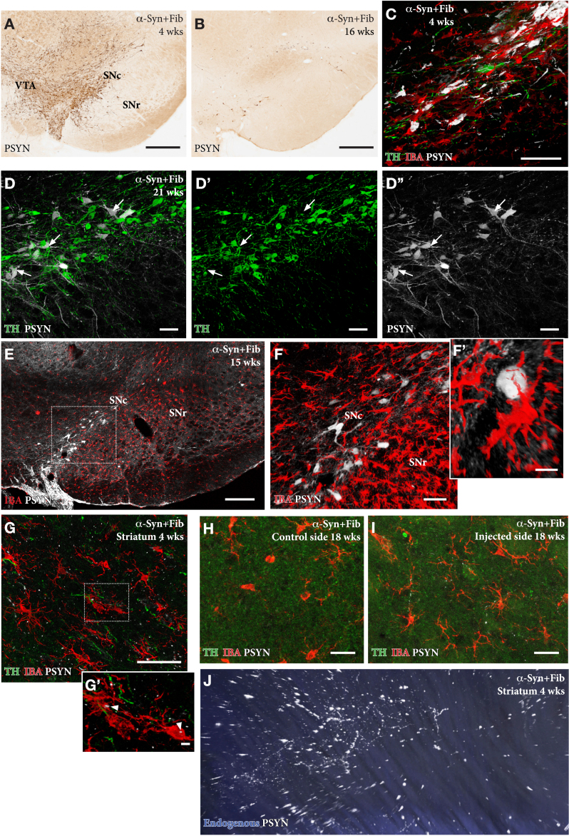
Fig. 4.
In the AAV/PFF treated rats prominent p-syn+pathology develops early, already at 4 weeks, in the DA neurons (A, C), as well as in their axons and terminals in the striatum, as illustrated in the Supplementary 3D movie captured by light-sheet microscopy (Supplementary Movie S1; still image in J). This is accompanied by an Iba1 + microglial response (F, G, H) and downregulation of TH (arrows in D-D''), that is maintained also at longer time-points. The p-syn pathology declines over time (B, E, F) which is in line with the progressive loss of the affected DA neurons. A notable feature of the microglial response is the appearance of p-syn+inclusions inside the Iba1 + microglia (arrow heads in G'). Scale bars: 500μm (A,B), 50μm (C,D,F,G,H,I), 200μm (E), 10μm (F',G').

