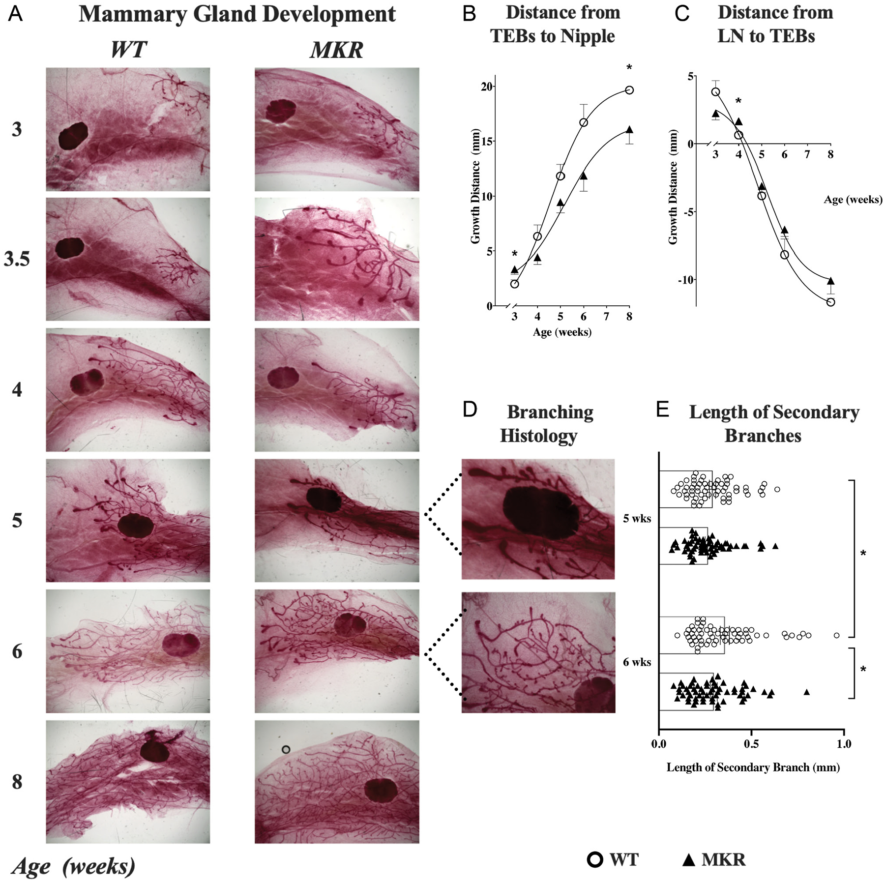
Figure 4.

Mammary gland development in MKR mice and WT mice. (A) Representative histology (1×) of the fourth mammary gland at 3, 3.5, 4, 5, 6, and 8 weeks of age from MKR and WT mice, showing the mammary ducts growing from their origination at the nipple toward then past the lymph node. The trajectory of gland development in relation to the nipple and lymph node was quantified in all MKR (▲, n = 30) and WT (O, n = 30) mice. (B) Ductal extension (mean ± s.e.m.) was measured as the distance between the nipple and the terminal end buds (TEBs). MKR mice show an initial accelerated growth spurt before 3.5 weeks of age, which then slows by 4 weeks to a final significant growth delay by 8 weeks. (C) Distance (mean ± s.e.m.) between the TEBs and the lymph node (LN). At 4 weeks of age, the growth trajectory of MKR and WT mammary glands cross over, as MKR growth falters relative to WT. (D) High powered view (4×) of branching pattern in MKR and WT mammary glands at 5 and 6 weeks of age, showing shortened length of branches and a structural abnormality of bulbs at the secondary branching points. (E) Length (mm) of secondary branches at 5 and 6 weeks of age in MKR (▲, n = 6) vs WT (O, n = 6), with 20 branches recorded per mouse.*P ≤ 0.05. A full color version of this figure is available at https://doi.org/10.1530/JOE-21-0447.
