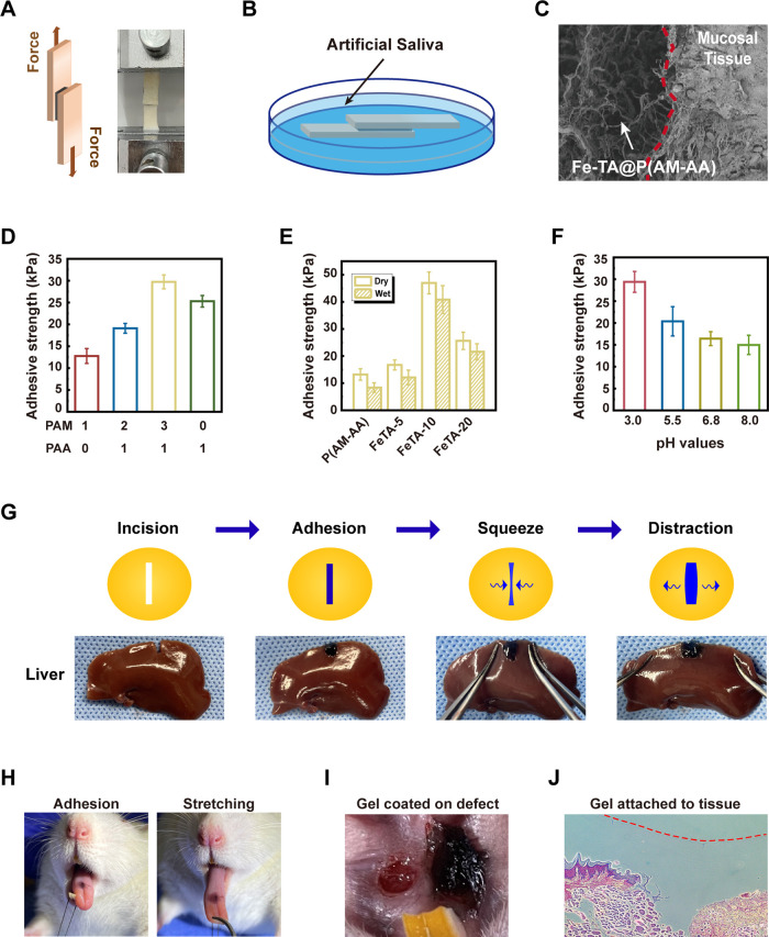
Figure 3.
Adhesive performance of the Fe-TA@P(AM-AA) hydrogel. Models of porcine skin adhesive strength test via standard lap shear measurements in both (A) dry and (B) wet environments. (C) SEM observation of the integration of the Fe-TA@P(AM-AA) hydrogel with rat oral mucosal tissues (red dashed line indicates the boundary). (D) Adhesive strength of hydrogels with different ratios of PAM and PAA. (E) Adhesive strength of composite hydrogels under dry and wet conditions. (F) Adhesive strength of composite hydrogels soaked in artificial saliva with pH values of 3.0, 5.5, 6.8, and 8.0. (G) Images of the adhesiveness of the Fe-TA@P(AM-AA) hydrogel to the incisions on fresh rat liver. (H) Images of the superior adhesion and deformation of the Fe-TA@P(AM-AA) gel on the wet rat tongue along with tongue movement. (I) Images of the Fe-TA@P(AM-AA) gel applied in oral wound areas of diabetic rats. (J) Histological sections of rat oral mucosa, verifying the integration of the Fe-TA@P(AM-AA) gel with the mucosal epithelium.

