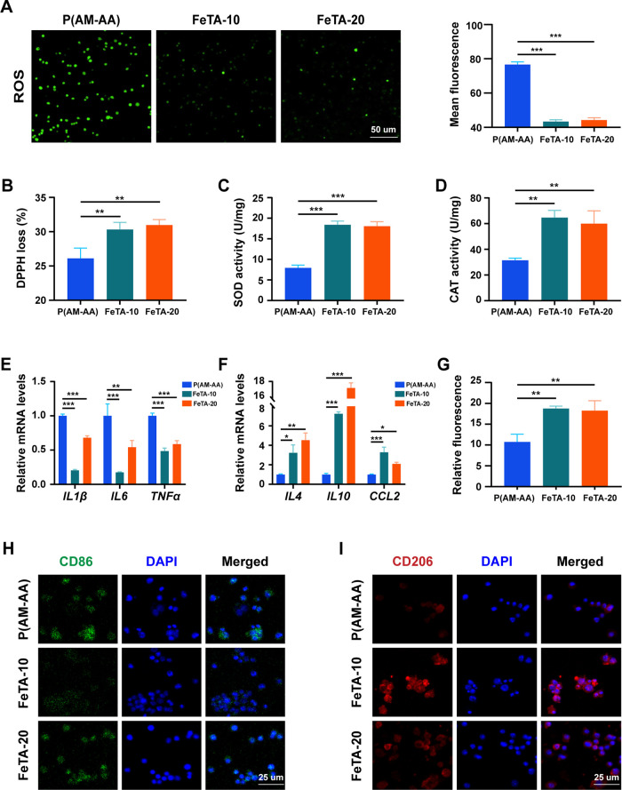
Figure 5.
Fe-TA@P(AM-AA) exhibits obvious antioxidant capacity and improves the inflammatory microenvironment under high-glucose conditions in vitro. (A) Intracellular reactive oxygen species (ROS) production of macrophages cocultured with different hydrogel extracts for 48 h. (B) Free radical scavenging test for different composite hydrogels in vitro by incubation with DPPH for 30 min. (C) Intracellular superoxide dismutase (SOD) activity of macrophages cocultured with different hydrogel extracts for 48 h. (D) Intracellular catalase (CAT) activity of macrophages cocultured with different hydrogel extracts under high-glucose conditions for 48 h. (E, F) RT-qPCR analysis of the expression levels of M1 macrophage markers (IL1β, IL6, and TNFα) and M2 macrophage markers (IL4, IL10, and CCL2) in THP-1 cells cocultured with hydrogel extracts under high-glucose conditions for 48 h. (G) Percentage of CD206+ M2 macrophages measured by flow cytometry after coculturing with hydrogel extracts under high-glucose conditions for 48 h. (H, I) Immunofluorescence of macrophage M1 and M2 polarization status induced by hydrogel extracts under high-glucose conditions in vitro.

