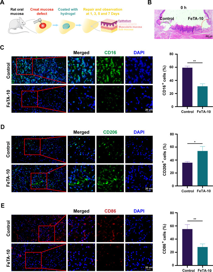
Figure 6.
Fe-TA@P(AM-AA) improves the inflammatory microenvironment of diabetic oral wounds in vivo. (A) Scheme for the creation and covering of a diabetic rat palatal mucosal defect model to assess the wound-healing potential of gels. (B) H&E staining showing the successful creation of mucosal defects of the same size in the epithelium and lamina propria (labeled with dashed arrows) on both sides of the palatal mucosa. (C) CD16-stained neutrophils in the defect regions, with higher-magnification images of the regions marked by red boxes shown on the right. The percentage of CD16-positive cells was measured. (D) CD206-stained M2 macrophages in the defect regions, with higher-magnification images of the regions marked by red boxes shown on the right. The percentage of CD206-positive cells was measured. (E) CD86-stained M1 macrophages in the defect regions, with higher-magnification images of the regions marked by red boxes shown on the right. The percentage of CD86-positive cells was measured.

