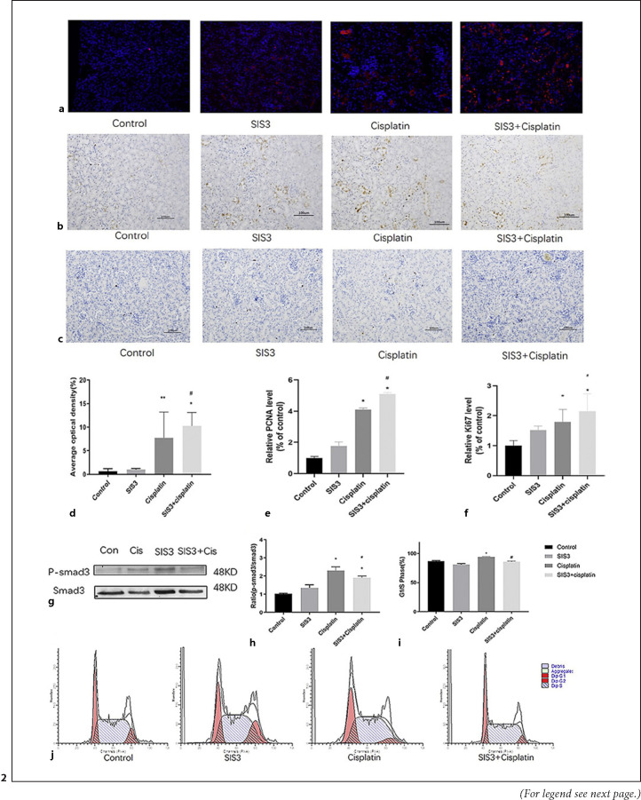
Fig. 2.
SIS3 promotes tubular epithelial cell regeneration in cisplatin-induced acute kidney injury. a BrdU staining shows SIS3 promoted the proliferation of tubules in cisplatin-induced AKI. b IHC shows SIS3 increased the expression of PCNA protein in cisplatin-induced AKI. c IHC shows SIS3 increased the expression of Ki67 protein in cisplatin-induced AKI. d–f Quantitative analysis of BrdU+, PCNA+, and Ki67 + mTECs. g Western blot shows SIS3 decreases the level of p-Smad3 protein in cisplatin-induced renal tubular epithelial cell. h Quantitative analysis of p-Smad3 by Western blot. i Quantitative analysis of the proportion of cells in G1/S phase by flow cytometry. j Flow cytometry analysis detects that SIS3 decreased the proportion of cisplatin-induced renal tubular epithelial cell in G1/S phase. Each bar represents the mean ± SEM. *p < 0.05, **p < 0.01, versus the control group. #p < 0.05, ##p < 0.01, versus the cisplatin group.

