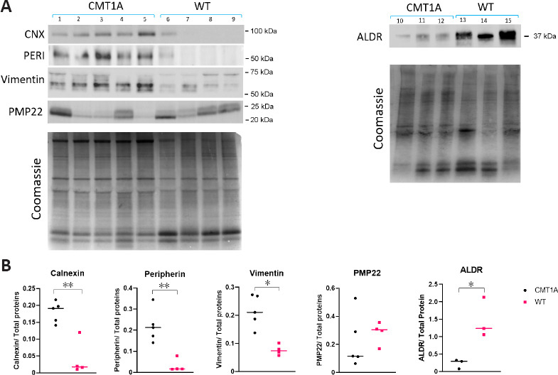
Figure 7.

Protein expression analysis.
(A) Western blot performed on sciatic nerve homogenates of CMT1A (n = 5) and WT (n = 4) rats aged 3 months. Calnexin (CNX), peripherin (PERI), vimentin, and peripheral myelin protein 22 (PMP22) staining were probed. For aldose reductase (ALDR), additional samples of the same age were used: CMT1A (n = 3) and WT (n = 3). As a loading control, the gel was stained by Coomassie staining. Shown is the Coomassie gel for PMP22 and vimentin blots, as well as the gel for ALDR blot. CNX and PERI were normalized on another blot. (B) Densitometric quantification of a shows a statistically significant high expression of CNX in CMT1A samples compared to WT samples (P < 0.01). Peripherin and vimentin showed overexpression in CMT1A samples in comparison to WT (P < 0.01 and P < 0.05, respectively). PMP22 quantification showed no statistical significance between CMT1A and WT. ALDR showed an underexpression in CMT1A samples (P < 0.05). The intensity of each sample was normalized by the intensity of total proteins per lane indicated by Coomassie staining. The median of values is shown. *P < 0.05, **P < 0.01 (Student’s t-test). CMT1A: Charcot-Marie-Tooth-1A; WT: wild-type.
