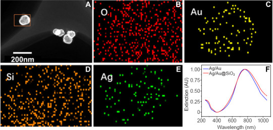
Figure 3.

Characterization of TPM4-targeted Ag/Au hollow nanoshells@SiO2 nanoparticles.
The prepared material showed a nanometer particle with a cage structure and diameter of approximately 80 nm. (A) Transmission electron microscopy image of Ag/Au@SiO2. Scale bar: 200 nm. (B–E) The corresponding element distribution mapping and energy dispersive spectrometer results. (F) The ultraviolet-visible absorption spectrum for Ag/Au and Ag/Au@SiO2 showed peaks at 808 nm. AU: Absorbance unit.
