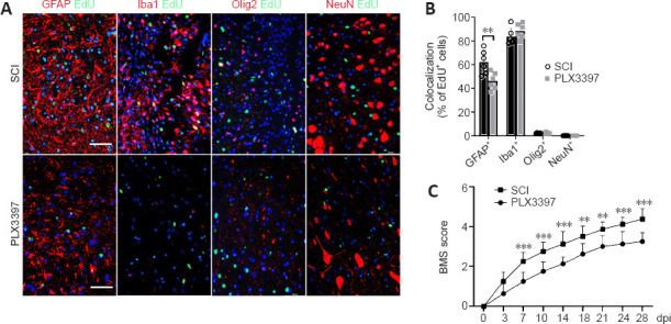
Figure 2.

Microglial depletion results in a reduced number of proliferating cells (especially astrocytes) in the spinal cord lesion area.
(A) Double immunofluorescence staining of EdU and indicated neural markers at the injury site at 7 days post-injury (dpi). EdU was predominantly colocalized with GFAP+ astrocytes and Iba1+ microglia/macrophages; only a small portion of Olig2+ cells were colabeled with EdU, and no EdU+/NeuN+ neurons were observed in each group. Scale bars: 50 μm. (B) Quantification of EdU incorporation in different cell markers, as determined by EdU+/GFAP+, EdU+/Iba1+, EdU+/Olig2+ and EdU+/NeuN+ cells. PLX3397 significantly reduced EdU incorporation in GFAP+ cells (unpaired t-test, n = 6). (C) The Basso Mouse Scale (BMS) score curve showed that the recovery of the SCI mice was significantly better than that of the PLX3397-treated mice from 7 dpi through the end of the experiment (two-way analysis of variance with Tukey’s post hoc test; n = 8). **P < 0.01, ***P < 0.001. Data are expressed as the mean ± SD. The experiments were repeated six times. EdU: 5-Ethynyl-2-deoxyuridine; GFAP: glial fibrillary acidic protein; Iba1: ionized calcium binding adaptor molecule 1; NeuN: neuronal nuclei; PLX3397: colony-stimulating factor 1 receptor inhibitor; SCI: spinal cord injury.
