Figure 4.

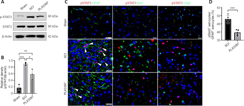
Microglial depletion inhibits phosphorylated STAT3 (pSTAT3) in astrocytes in spinal cord tissue.
(A) Western blotting bands of STAT3 and pSTAT3 at 7 post-injury (dpi). (B) Quantification of pSTAT3 (one-way analysis of variance with Tukey’s post hoc test, n = 3). Data are expressed as the mean ± SD. (C) Double immunofluorescence staining of pSTAT3 with various markers at 7 dpi. Arrowheads indicate double-stained cells. Scale bars: 50 μm. (D) Colony-stimulating factor 1 receptor inhibitor PLX3397 treatment significantly decreased the proportion of pSTAT3+ astrocytes (unpaired t-test, n = 5). *P < 0.05, **P < 0.01, ***P < 0.001. Western blot experiments were repeated three times. Immunofluorescence staining was repeated five times. GFAP: Glial fibrillary acidic protein; Iba1: ionized calcium binding adaptor molecule 1; SCI: spinal cord injury; STAT3: signal transducers and activators of transcription 3.
