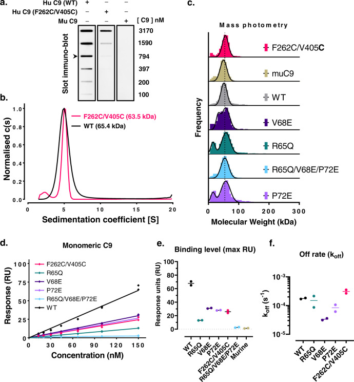
Fig. 4. Monomeric C9 is weakly recognised by aE11.
a A concentration series of human, murine, and disulphide trapped C9 variants in the monomeric state detected by aE11 IgG in a slot immunoblot assay. An arrow shows the approximate physiological concentration of monomeric C9 (900 ± 200 nM)59,60. b Analytical ultracentrifugation of wild type and disulphide-trapped C9 at micromolar concentration shows only monomeric C9 species. c Mass photometry measurements of the molecular mass of C9 variants corroborates AUC measurements (n = 2). Lower molecular weight peaks (~10 kDa) of R65Q and P72E variants correspond to a known contaminant. d Maximal SPR response of aE11 IgG binding versus concentration of monomeric C9 and variants (n = 2). e Maximal binding of aE11 IgG to monomeric C9 at 150 nM (n = 2). f Kinetic fit of off-rate for one-to-one binding of aE11 IgG to monomeric C9 (n = 2). Individual data points and their means are plotted.

