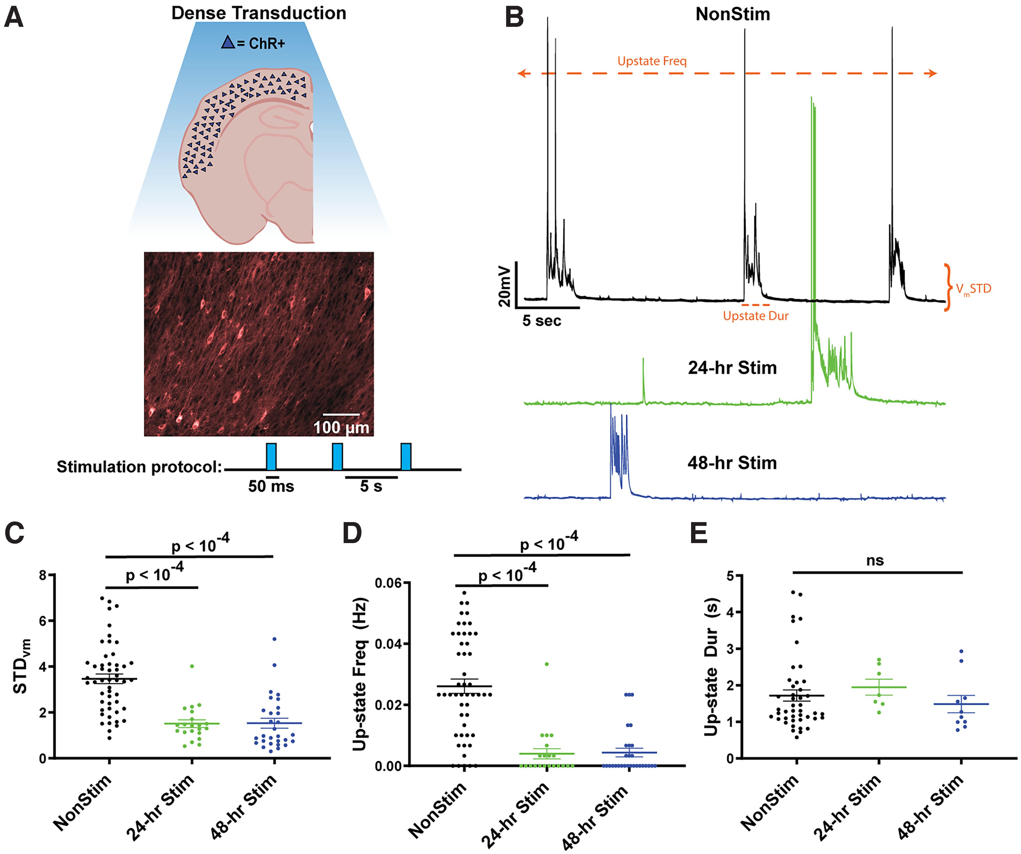
Figure 1.

Spontaneous Up-state frequency is reduced in densely transduced cortical slices following 24 or 48 h of stimulation. A, Schematic of densely transduced cortical circuits in organotypic slice cultures (top) and image from auditory cortex densely transduced with AAV9-CaMKIIα-hChR2(H134R)-mCherry and chronic optogenetic stimulation paradigm (bottom). B, Example traces of spontaneous Up-states in Pyramidal neurons from unstimulated (black), 24 h stimulated (green), and 48 h stimulated (blue) slices. Up-states were rarely observed in the 24 and 48 h stimulated slices. Orange annotations represent the three quantitative measures of spontaneous activity shown in C–E. C, The SD of membrane voltage was significantly decreased by both 24 and 48 h of stimulation. STDVm was calculated over a 5 min period of spontaneous activity in Pyr neurons. D, Spontaneous Up-state frequency was significantly decreased by both 24 and 48 h of stimulation. E, Although Up-state frequency was decreased by stimulation, when Up-states occurred, on average, they were of the same duration.
