
Figure 2.
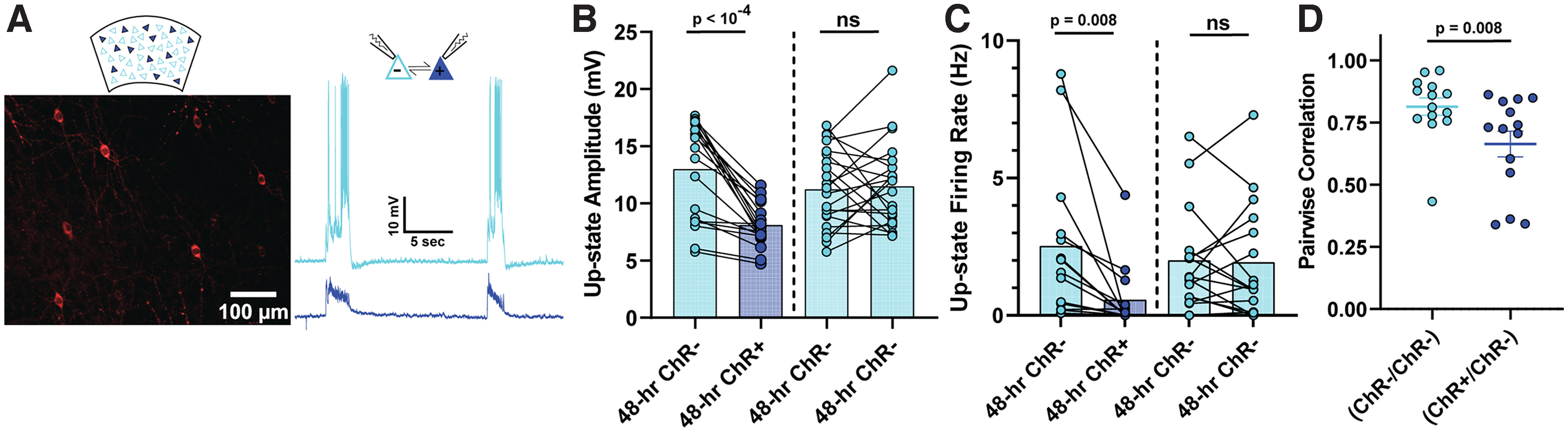
Pairwise differences in Up-state amplitude, firing rate, and voltage correlation between stimulated and nonstimulated pyramidal neurons in sparsely transduced slices. A, Example of cortical pyramidal neurons sparsely transduced with AAV9-CaMKIIα-Cre and EF1a-DIO-hChR2(H134R)-mCherry (left), and sample paired recordings of ChR+ and ChR– neurons (right). B, Spontaneous Up-state amplitude was significantly reduced in ChR+ compared with ChR– pyramidal neurons. Up-state amplitude was not significantly different between simultaneously recorded ChR+ pyramidal neurons grouped according to their resting membrane potential (ChR– pyramidal neurons with the lower resting membrane potential of the pair was plotted on the left). C, Spontaneous Up-state firing rate was significantly reduced in ChR+ versus ChR– pyramidal neurons. Up-state firing rate was not significantly different between simultaneously recorded ChR– pyramidal neurons grouped according to their resting membrane potential. D, The correlation between the Up-state voltage dynamics of ChR+ and ChR– neurons was significantly less than ChR– and ChR– pairs, indicating a decorrelation between the shared inputs to the ChR+ and ChR– subpopulations.