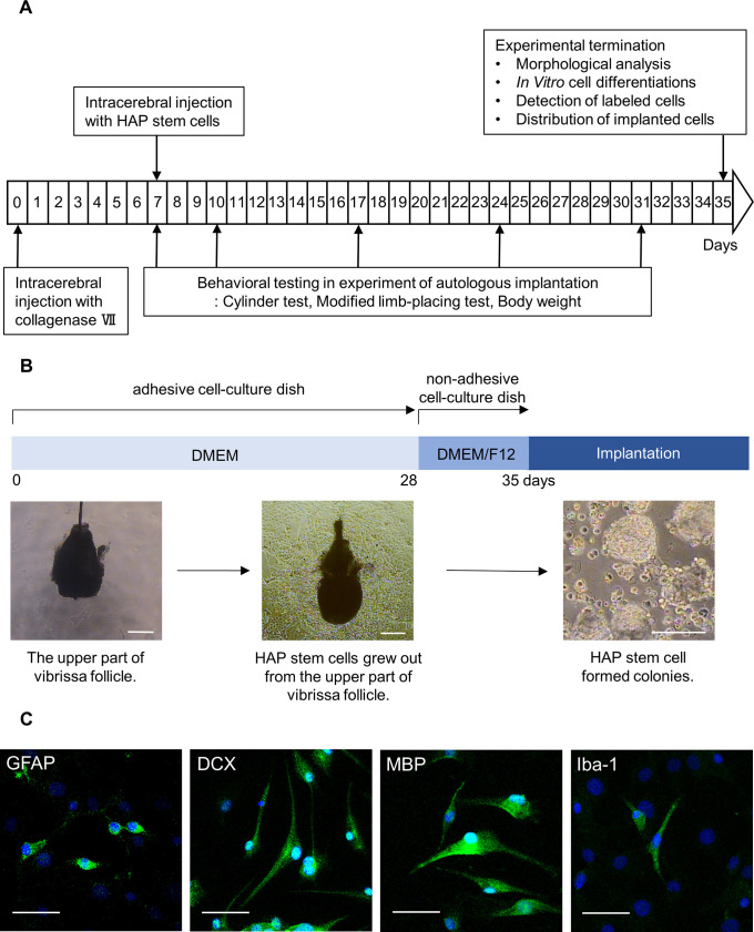
Fig 1. Experimental flow diagram and culture of HAP stem cells.
(B) Procedure for culture and colonization of HAP stem cells. Bar = 200 μm. (C) Immunofluorescence staining shows that the cultured HAP stem cell colonies differentiated to GFAP-positive astrocytes, DCX-positive neurons, MBP-positive oligodendrocytes and Iba-1-positive microglia on Lab-Tek chamber slides. Green = GFAP, DCX, MBP or Iba-1; Blue = DAPI. Bar = 50 μm.

