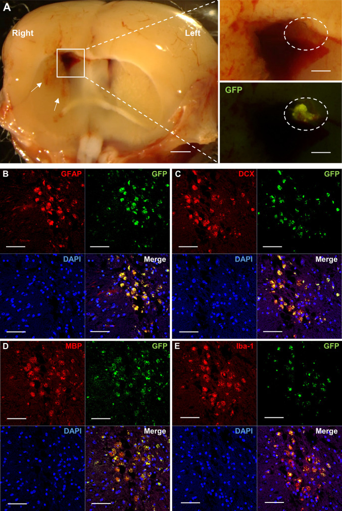
Fig 2. Implanted HAP stem cells repair the ICH site in nude mice.
(A) Stereomicroscopy shows that GFP-expressing HAP stem cells repaired the ICH lesion. White arrow indicates hemorrhage in the striatum. White square line indicates hematoma in the brain. White dotted line indicates transplanted HAP stem cells from GFP mice in the hematoma. Left panel = low-magnification of coronal section at ICH. Bar = 1mm. Right panel = high-magnification of white square line area. Bar = 500 μm. (B-E) Immunofluorescence staining shows that the implanted HAP stem cells differentiated to astrocytes (B), neurons (C), oligodendrocytes (D) and microglia (E) in the ICH area. Red = GFAP (B), DCX (C), MBP (D) or Iba-1 (E); Green = GFP (B-E); Blue = DAPI (B-E); Merged (B-E). Bar = 50 μm. Insets show a higher magnification; Bar = 25 μm. All images show coronal sections of the brain.

