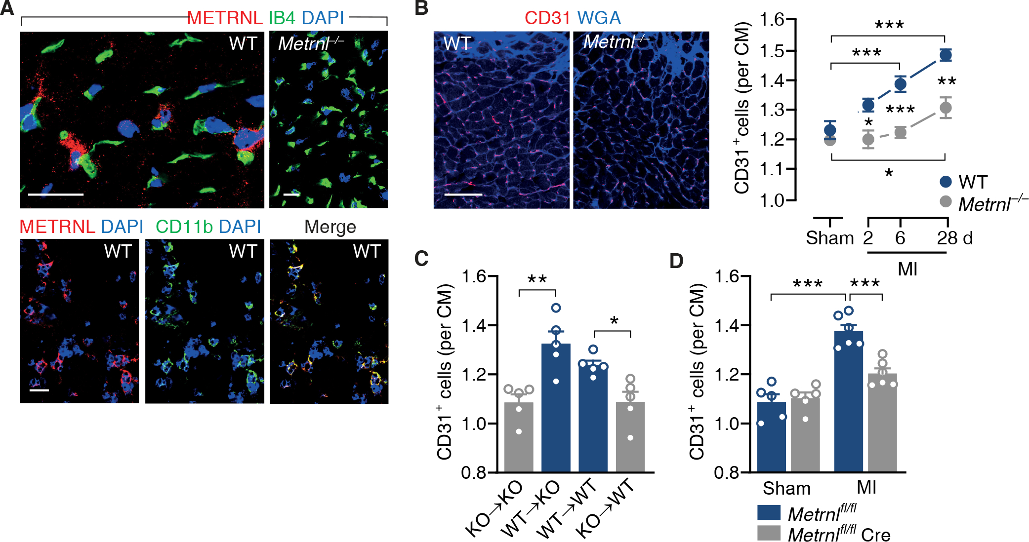
Fig. 1. Myeloid cell-derived METRNL promotes angiogenesis after myocardial infarction.

(A) Confocal immunofluorescence microscopy images taken from the infarct border zone 3 days after myocardial infarction (MI) in wild-type (WT) or M etrnl−/− mice. Sections were stained with DAPI, fluorescein-labeled isolectin B4 (IB4), and antibodies against METRNL and CD11b. 92 ± 5% of the METRNL-expressing cells co-expressed CD11b (data from 4 mice). Scale bars, 25 μm. (B) WT and Metrnl−/− mice underwent sham or MI surgery and were followed for 6 (sham) or up to 28 days (MI). Fluorescent images depict CD31+ endothelial cells in the infarct border zone on day 28. Extracellular matrix and cardiac myocyte (CM) borders are highlighted by WGA staining. Scale bar, 50 μm. Summary data are from 5 mice per group. One-way ANOVA with Dunnett test (MI vs. same genotype sham), independent-samples t test (WT vs. Metrnl−/−). (C) Bone marrow cells from Metrnl−/− (knockout, KO) or WT mice were transplanted into (→) lethally irradiated KO or WT recipients. MI was induced after bone marrow reconstitution and CD31+ capillary density in the infarct border zone was determined on day 28. Independent-samples t test. D) CD31+ capillary density in the infarct border zone in Metrnlfl/fl mice and Metrnlfl/fl LysMCre/+ (Metrnlfl/fl Cre) mice on day 28. Two-way ANOVA with Tukey test. (B to D) Data are means ± SEM. *P < 0.05, **P < 0.01, ***P < 0.001.
