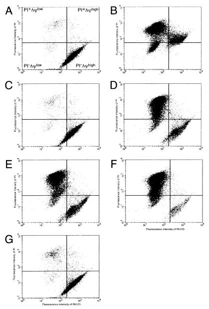
FIG. 1.
Flow cytometric analysis of HL-60 cells incubated with or without 50 μg of protein of the extracts from various bacteria per ml. Panels: A, PBS; B, adriamycin (10 μM); C, E. coli; D, B. forsythus; E, A. actinomycetemcomitans serotype a; F, A. actinomycetemcomitans serotype b; G, A. actinomycetemcomitans serotype c. The extent of cell death was assessed by measuring fluorescence intensity using a FACScalibur flow cytometer after staining with PI and Rh123. All analyses were performed in duplicate experiments.

