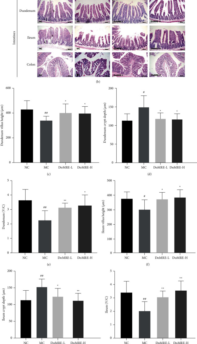
Figure 5.

Effect of DoMRE on histopathological changes in the renal and intestine. (a) Renal tissues were stained with H&E at a magnification of 200x: (A) glomerulus significantly atrophy; (B) glomerular rupture; (C) the renal tubules swollen and deformed. (b) The duodenum, ileum, and colon were stained with H&E staining to observe crypt (A) and villus (B) at 200x. (c–h) The villus height (V), crypt depth (C), and V/C ratio in duodenum and ileum. IOD: integrated optical density; NC: normal control group; MC: model control group; DoMRE-L: low dose of DoMRE group; DoMRE-H: high dose of DoMRE group. The data were expressed as mean ± SD of 8 rats in each group. #P < 0.05; ##P < 0.01, compared with NC group; ∗P < 0.05; ∗∗P < 0.01, compared with MC group.
