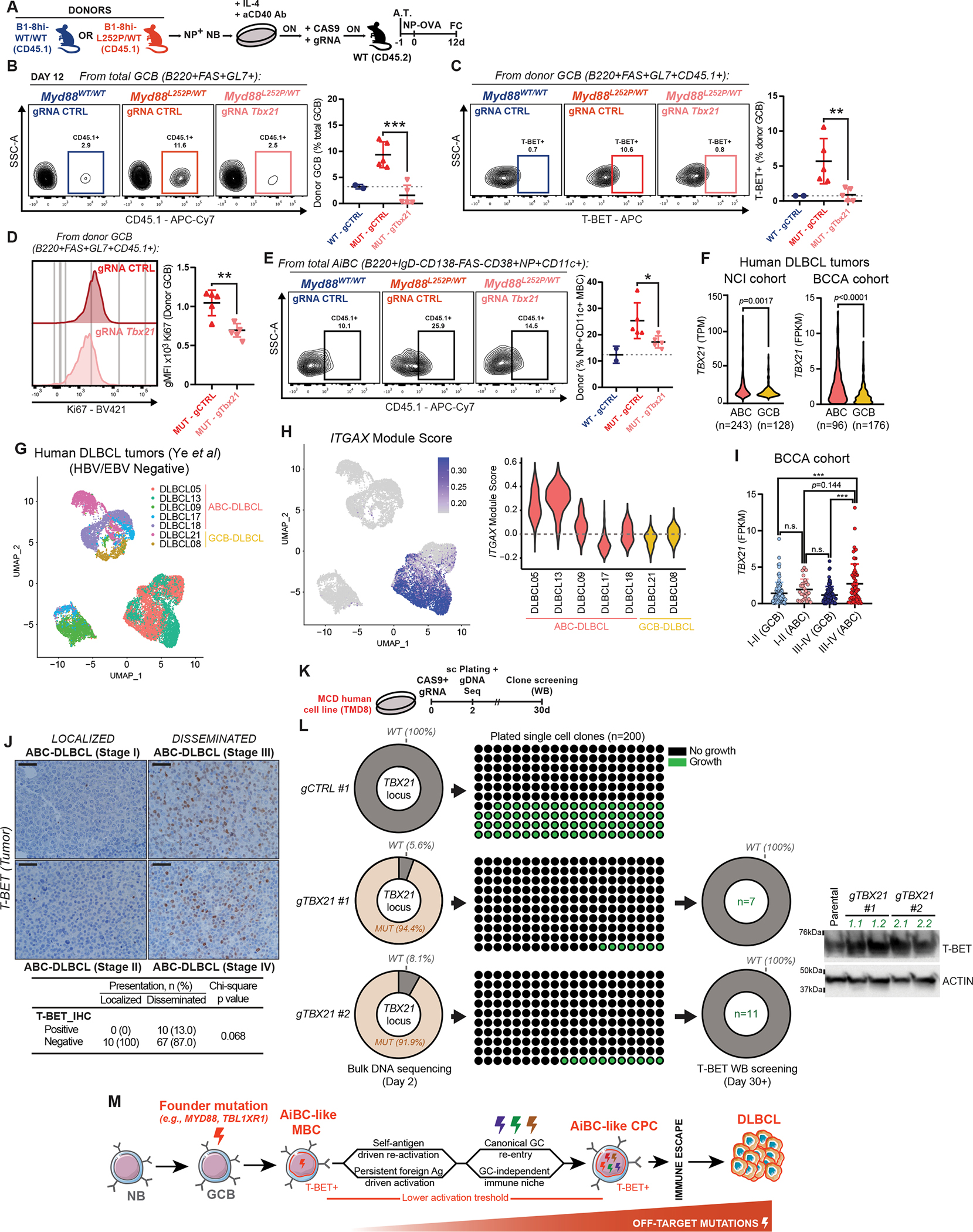
Figure 7. T-BET supports the fitness of MYD88-mutant B-cells.

A, Experimental scheme for B-D. ON = Overnight; A.T. = Adoptive transfer.
B, FC profiling of donor B-cell contribution to total GCB.
C, FC analysis of T-BET+ donor-derived GCB.
D, FC analysis of KI67 expression in donor-derived GCB.
E, FC analysis of donor B-cell contribution to AiBC-like MBC.
F, RNA-Seq-based TBX21 expression in primary specimens from (left) NCI (47) or (right) BCCA (48,49) cohorts.
G, uMAP depiction of single-cell RNA-Seq data from EBV/HBV-negative DLBCL tumors (50).
H, Relative expression of the ITGAX module among specimens in (G).
I, RNA-Seq-based TBX21 expression in primary human DLBCL specimens.
J, Representative images and quantification of T-BET IHC in specimens from the BCCA cohort (52). Scale = 20μm.
K, Experimental scheme for (L).
L, (Left) Penetrance of targeted genomic alterations, at time of cell plating. (Center) Number of detectable clonal outgrows 30 days after plating. (Right) WB-based T-BET expression in clonal outgrows. Representative blots for 2 clones per gRNA.
M, Schematic representation of the proposed transformation model.
Values represent mean ± SEM. P-values calculated using unpaired two-tailed Student’s t-test (B-F), or one-way ANOVA with Tukey’s post-test (I).
